Beverly Hills, California, dentist explains the benefits of dental imaging for diagnostics and planning

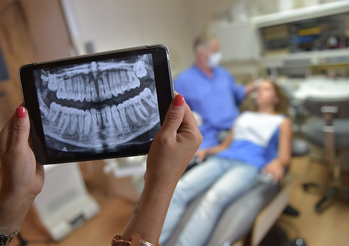
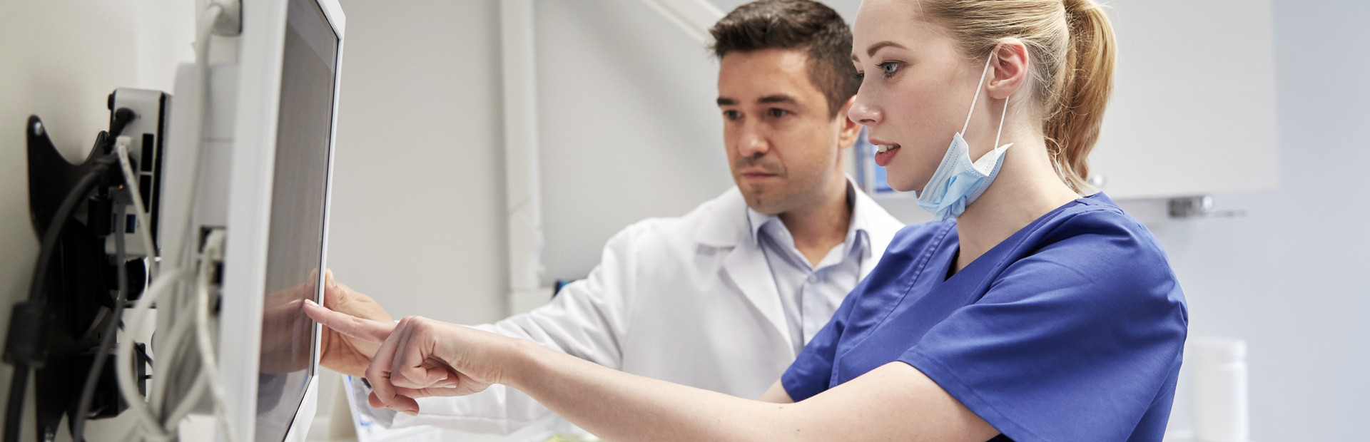
Dental imaging, or dental radiography, can help a dentist dramatically. These images are essential to preventive diagnostics, and planning of dental issues. Dentists and dental hygienists use the information found in radiographs to detect dental abnormalities that cannot be seen with the eye and hide underneath the gums. This imaging is also used to develop a treatment plan for a patient or determine the best place to put a dental implant for restoring the smile. It can also be used for those considering bone grafting.

What is dental imaging used for?

In addition to a physical evaluation of the oral cavity during routine appointments, dental imaging can take diagnostics one step further. They are used to reveal:

  • Bone loss
  • Cysts/abscesses
  • Tooth decay
  • Tumors
  • Developmental abnormalities
  • Problems inside of a tooth
  • Problems underneath the gum line.

With dental imaging, many conditions are spotted at an early stage, which can dramatically improve a patient’s outcome. Early detection of many conditions can save patients time, money, and even teeth.

Is dental imaging safe?

Many patients are worried about the radiation that comes from dental imaging and ask our team whether it’s safe. Patients need to understand that natural radiation occurs every day in our environment, and radiation exposure during dental imaging is comparable to a patient’s daily exposure during their everyday lives. Dental imaging produces such a low level of radiation that they are considered safe. Even so, our dental team takes proper precautions to reduce exposure by using lead apron shields and faster film imaging.

How often do I need to undergo dental imaging?

Your dentist and dental hygienist will make a recommendation during your appointment. New patients may have dental imaging done during their first appointment, but not at consecutive visits unless there is a specific problem. Patients coming into the office for toothaches or other complaints may have dental imaging done to look for the root of the problem, which may be beneath the gums or inside of a tooth.

Dental Imaging FAQs

Why do I need dental imaging?

Many dental problems develop below the surface where we can’t see them with a visual exam. Imaging helps us detect abscesses, bone loss, impaction, cysts, and root issues before they become painful emergencies. Early detection means simpler treatment and better long-term outcomes.

What should I expect when I get dental X-rays?

Getting dental X-rays is quick, comfortable, and non-invasive. We’ll ask you to bite down on a thick paper tab for a few seconds while we take the X-rays. There’s no lingering discomfort, and the procedure is finished in minutes. Most patients are surprised by how easy it feels.

How long should I expect the results?

With modern imaging, results are available in real time. Your doctor can review the images on a chairside screen and explain what they mean in real time. There’s no waiting period or separate appointment needed to discuss findings. Quick results ensure faster turnaround, resulting in fewer interruptions in your everyday life at home, work, or school.

How is 3D scanning different from standard X-rays?

Traditional X-rays provide flat, two-dimensional images. 3D scanning creates a detailed, 360-degree view of your teeth, jawbone, and surrounding structures. 3D imaging is especially important in planning dental implants and endodontic treatments, where precision is essential to achieve the best possible outcomes.

How does my local endodontist in Beverly Hills use dental imaging?

Endodontic imaging guides every stage of our endodontic treatments—there’s no room for guesswork. X-ray images and 3D scans help map canal anatomy, confirm infections, guide treatment, and monitor post-op healing. Dr. Fanny Yacaman uses advanced imaging to treat teeth conservatively while maximizing long-term success.

What is the recommended frequency for receiving dental X-rays?

Frequency depends on your age, oral health history, and individual risk. We recommend X-rays every 1-2 years for patients without oral health risks. However, you may need X-rays more frequently if you are vulnerable to cavities, gum disease, and other issues. Children also need X-rays more often than adults to monitor tooth and jaw development. Our office follows evidence-based guidelines to recommend the frequency of X-rays.

Are dental X-rays safe?

We get this question a lot, and we are excited to answer it. Modern digital X-rays emit extremely low radiation levels, typically 80-90% lower than those of older film-based systems. Still, we use lead aprons and other protective gear to minimize unnecessary exposure. The benefits of digital radiographs far outweigh the minimal risks involved.

Should I get dental X-rays if I am pregnant?

Routine X-rays are often postponed during pregnancy, especially during the first semester. When imaging is required, such as during emergencies, we use protective shielding to keep exposure as low as possible. Always let us know if you are pregnant or think you might be, so we can tailor your procedure to maximize your safety and that of the unborn baby.

Can I breastfeed after a dental X-ray?

Yes. Dental X-rays do not affect breast milk. There is no need to pause breastfeeding after imaging. If you still have concerns, talk to our dentist about your specific condition.

Where can I find a dentist in Beverly Hills, CA who uses modern technology?

Beverly Hills Advanced Specialties of Dentistry has invested in technology that makes a real difference in your dental care. Modern technology promotes precise diagnosis, targeted treatment, and a smoother patient experience. Call (310) 861-3955 or (310) 878-6455 and experience redefined care with our advanced technology.

Schedule an appointment today to have a full evaluation performed with dental imaging

Contact Beverly Hills Advanced Specialties of Dentistry in Beverly Hills, CA, today at (310) 878-6455. Our dentists, including Drs. Frank Vidjak and Fanny Yacaman are committed to helping in early diagnosis of dental problems with dental imaging solutions.

The skilled doctors at our practice are Frank M. A. Vidjak, DDS, MSEd and Fanny Yacaman, DDS, MSEd, MS.


Doctors Bio Image - Dr.Frank Vidjak
Dr. Vidjak has been a prosthodontist for over two decades, with his private practice in Beverly Hills from 1989. A USC School of Dentistry graduate, he completed his specialty training in prosthodontics and an MS in Medical Education at USC where he also taught the School of Dentistry. He was its Clinical Director of the Advanced Prosthodontic Program and International Student Program too.


Doctors Bio Image - Dr. Fanny Yacaman
Dr. Yacaman received her DDS from the University Technologica of Mexico and served the clinical faculty in Endodontics at Unitec. She practiced Endodontics in Mexico City for over 10 years. She has an advanced specialty certificate in Advanced Microscopic Endodontics and Microsurgery and a Master of Science in Medical Education from the USC and has been a Clinical Faculty member at the UCLA School of Dentistry Department of Endodontics for more than 15 years. She specializes in Root Resorption treatment, having researched on External Root Resorption for over 15 years for a Master’s in Craniofacial Molecular Biology at USC’s Center for Craniofacial Molecular Biology. She is one of four Endodontists in United States that can treat Root Resorption. She is a local and international lecturer in Endodontics and Microsurgery and has received many awards and has published articles in the area.